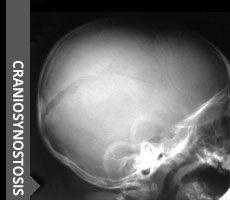
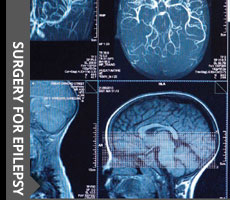
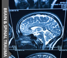
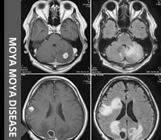
DISEASE &
TREATMENTS
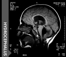
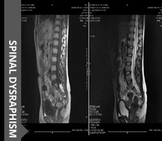
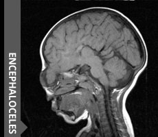
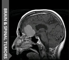
Click to know more about neurological disorders and diseases and it's treatment.
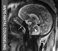
15 month girl 4 months back had sudden squinting recovered almost completely in 30 days time head circumference at 95th percentile AF still open full pusatile no papilledema no Headache no vomiting MRI Brain done
Its Suprasellar Archnoid Cyst causing secondary hydrocephalus
I did endoscopic septostomy followed by ventriculocystostomy and then cystostomy into basilar cistern
Endoscopy video , first we enter the right frontal horn and see the arachnoid cyst occupying whole of the right fornamen on monro and stretching the fornix , the surface that is visble is arachnoid cyst and upward elevated floor of third ventricle , the endoscope is turned medialy to do spetostomy first and enter opposite ventricle , then the cyst is fenestarated with two holes and the holes are communicated with scissor, then the cyst is entered and one can see the basilar artery both PCA and left 3 rd and 4 th nerve and at the base of the cyst just anterior to basilar artery another fenestration is done
Ct cisternography done after 72 hrs of surgery
First image is before the dye injection as base line
Second image is 5 minutes after dye injection
Third one is after 30 minutes
One can see dye in the basilar cistern and also 4th ventricle
There r high chances of stenosis we prevent by three steps 1 using monopolar as minimum and make sharp cut on the archnoid cyst wall with scissor
2 the basilar cistern perforation is done bluntly without cautery and pressure drives the CSF across the stoma and keeps it open
3 i put reservior with ventricular end few holes in the cyst and few holes in the ventricle across the stoma site so that it acts as conduit internally even if the cyst wall collapses and closes
Click to know more about neurological disorders and diseases and it's treatment.
© 2016 ChildNeuroSurgeon.com. All rights reserved | Design by MHI