DISEASE &
TREATMENTS
Click to know more about neurological disorders and diseases and it's treatment.
Epilepsy
Epilepsy is an intermittent derangement of the nervous system due to a sudden, excessive, disorderly electrical discharge of cerebral neurons. The term epilepsy is derived from the Greek word epilamvanein .Prevalence rate of epilepsy in India is 5.59 per 1000 population with no gender or geographical difference. More than two-thirds of all epilepsy begins in childhood, when seizures can have drastic and devastating results on the child's psyche and development. Recent advances over the past decade in the understanding of pathogenesis mechanisms and in the diagnosis of epilepsy have had a significant impact on every aspect of epilepsy management including surgical management of epilepsy.
Surgical treatment is highly effective in the treatment of many of the epilepsies that have not responded to medical therapy. Nearly 40 percent of children with partial epilepsy are candidates for surgical therapy.
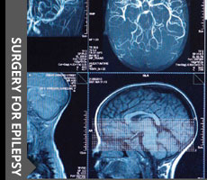
I work in close association with a team. Our Pediatric Epilepsy Surgery team includes Pediatric Neurologists, Pediatric Neurosurgeons, Neuropsychologists and Neuroradiologist. Children with medically intractable epilepsy are extensive evaluations include video-EEG, Neuroimaging like MRI. and Neuropsychological assessments. We conduct monthly meetings to discuss pediatric epilepsy cases in our forum to provide the best surgical management and plan [tailored to the individual’s case]and care. The department is experienced in performing temporal lobectomy, lesionectomy, corpus callostomy and hemispherectomies and placement of vagal nerve stimulator. I also conduct Awake craniotomy when appropriate.
Why epilepsy surgery?
AED is main stay in the treatment. The untoward cognitive side effects of AEDs coupled with the long-term effects of recurrent seizures, reduced quality of life and increased co-morbidities for the majority of patients. So in children with epilepsy where surgical option is available, more and more children are recruited for epilepsy surgery.
Patients with unsatisfactory seizure control often seek alternative care. The number or severity of the seizures may be unacceptable to the patient, family, or treating physician. Other reasons for referral for epilepsy surgery include the results of diagnostic tests that may show a structural focal brain lesion, unsatisfactory psychosocial adaptation due to poor seizure control, unacceptable sedation, or other drug side effects.
The morbidity and mortality of seizures include accidental injury; cognitive decline; sudden death; and psychological, social, and vocational impairment. Accidental injuries commonly include fractures, burns, dental injuries, lacerations, and head injuries. Cognitive decline over time has been demonstrated to occur in patients with certain epilepsy syndromes who have recurrent convulsive seizures or episodes of status epilepticus. Both depression and anxiety are very common among patients with medically refractory epilepsy. Vocational issues include inability to be employed or, if employed, underemployment.Prolonged anticonvulsant drug therapy also affects cognition, memory, behaviorand motor developmentin substantial number of children.
Type of surgery
Epilepsy surgery may be divided into two major categories: Resective and Functional. The aim of Functional surgery is to decrease seizure frequency.
Resective surgeries
The aim of resective surgery is to remove the epileptogenic zone and render the patient seizure free. Surgeries done are:
1. Anteromedial Temporal Resection [AMTR],
2. Selective Amygdylohippocapectomy
3. Lesionectomy [Temporal and Extra-temporal]
Anteromedial Temporal Resection [AMTR]
AMTR comprises excision of mesial temporal structure (including the amygdala, hippocampal head and body, uncus, entrorhinal region, and the parahippocampalgyrus), and a variable portion of the tip of the temporal lobe. The greatest emphasis is on AMTR because this is the most commonly performed surgery and has best results
Selective Amygdylohippocapectomy [SAH]
In SAH, only amygdala and hippocampus is excised leaving the neocortex intact. Seizure outcome after SAH to be similar but there better neuropsychological outcome in SAH.SAH is technically difficult, time consuming, and at times, associated with some degree of cerebral swelling due to dividing several venous and arterial adhesions SAH should be reserved to resection on the dominant hemisphere.
Lesionectomy [Temporal and Extra-temporal]
With advancement in Neuroimaging, highly epileptogenic and resectable cortically-based lesions, are identified e.g. cavernoma, focal areas of cortical dysplasia, and indolent tumors such as Dysembryoplastic Neuroepithelial tumor [DNET], low grade astrocytoma, Ganglioglioma. These lesions can be safely resected. The extent of perilesional resection can be better determined by neuronavigation and intra-operative electrocorticography. The success epilepsy surgery depends on the complete resection of the epileptogenic zone
Functional epilepsy surgery
The objective in functional epilepsy surgery is to palliate rather than to cure the epilepsy. Functional procedures should only be considered once resective surgery has been deemed inappropriate, or to carry too great a risk. Functional procedures are
1. Corpus Callostomy
2. Hemispherectomy and Functional Hemispherotomy
3. Multiple Subpial Transection[MST]
4. Vagal nerve stimulation[VNS]
Corpus callostomy
The corpus callostomy is primarily done for atonic drop attacks, although it has been used to good effect in other epilepsy types and syndromes. The major risk with corpus callosotomy is that immediate or delayed disconnection syndrome (i.e., Mutism, left-sided apraxia that resembles hemiparesis, bilateral frontal lobe reflexes). In order to prevent or minimize the risk of a disconnection syndrome the callostomy is carried out in two stages, with the anterior two-thirds of the corpus callosum being divided at the first operation and the posterior third divided later. I also do a single-stage complete callosotomy whenever appropriate.
Hemispherectomy and Functional Hemispherotomy
Hemispherectomy and Functional hemispherotomy operation consists of disconnecting the abnormal epilepsy discharging hemisphereof brain from the normal hemisphere .The success of operation depends on the underlying pathology, with excellent outcomes expected for pathologies such as Rasmussen’s encephalitis and focal infarcts, and a poorer outcome expected in patients with hemimegalencephaly and cortical dysplasia.
Multiple Subpial Transections [MST]
Neocortex is organized in functional columnar units; right-angle cuts to the pial surface would not disrupt cortex-subcortical input-output interactions. Theoretically, if this intracortical synchronization can be disrupted by placing parallel slices through cortex is to permanently disrupt side-to-side intracortical synchronizing neural networks. The MST technique theoretically is ideal for treating epileptogenic involving the eloquent areas of the brain while preserving intrinsic cortical function.
VNS Vagal nerve stimulation
Vagal nerve stimulation was introduced in 1988. This technique involves intermittent stimulation of the cervical portion of the left vagus nerve by implanted electrodes connected to a subcutaneous generator. The mode of action is not entirely understood but is believed to involve inhibition of the brainstem and other subcortical structures. VNS indicated in uncontrolled epilepsy with Neuroimaging shows no structural lesion. VNS is well tolerated rare Side effects include hoarseness of voice, throat discomfort, and cough, which occur more commonly with a higher frequency of stimulation disadvantage is the huge cost factorin India.
Invasive Monitoring
Invasive monitoring is indicated when imaging does not conclude with the lesion and direct stimulation of the lesion is essential. Following are the techniques:
Intraoperative ECoG and Strip Electrodes: Strip electrodes are used most often to lateralize the side of seizure onset in frontal and temporal lobe epilepsy, but they may also be used to obtain survey studies over cortical surfaces of the brain.
Subdural Grid Electrodes: Subdural grid consists of electrodes grids with 5-8 rows (20-64 contacts) to maximize coverage over the craniotomy site inserted after craniotomySubdural. Cortical mapping helps in defining the epileptogenic cortex and also determine the language, motor, and sensory areas to mark the functional cortex.
Depth Electrodes: Depth electrodes are used most commonly for recording from the hippocampus and amygdala that is placed with neuronavigation
With advancement in technology and awareness among the pediatrician and pediatricneurologist in past decade there has been dramatic increase in the number of surgical procedures for epilepsy.
Click to know more about neurological disorders and diseases and it's treatment.
© 2016 ChildNeuroSurgeon.com. All rights reserved | Design by MHI